







Glaucoma & Retina
Supervised by Dr. Ankita Patil
Glaucoma, OCT & Retina Diagnostics
Detecting silent vision loss early through advanced visual field perimetry and retinal imaging.
Cashless TPA Insurances
0% EMI Options
Top Network Hospitals
Diagnostics
Humphrey Field + OCT
Laser Care
Nd:YAG Outpatient
Retina Review
Dilated Fundus Biomicroscopy
Target
Lifetime Vision Preservation

Clinical Treatment Breakdown
Evidence-based medical care tailored for precision, comfort, and fast functional recovery.
Conditions Treated
- Primary Open-Angle & Normal-Tension Glaucoma
- Angle-Closure Glaucoma & narrow anterior chamber angles
- Ocular Hypertension with elevated intraocular pressure (IOP)
- Diabetic Retinopathy (Microaneurysms, macular edema)
- Hypertensive Retinopathy and vascular occlusions
Treatment Approach
-
Humphrey Visual Field Perimetry: Gold-standard automated computerized mapping detecting subtle peripheral vision blind spots before you notice them.
-
Optical Coherence Tomography (OCT): High-resolution cross-sectional retinal and optic nerve fiber layer (RNFL) thickness analysis at micron resolution.
-
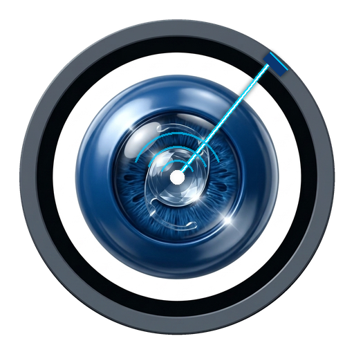
YAG Peripheral Iridotomy: Pain-free outpatient laser procedure creating a microscopic bypass channel to prevent acute angle-closure glaucoma attacks.
Recovery Milestones
Initial Workup
Complete tonometry, pachymetry, gonioscopy, and visual field map completed in a single 45-minute visit.
Target Pressure
Custom anti-glaucoma medication titration to achieve safe individualized target eye pressure.
Every 6 Months
Structured visual field and RNFL tracking to guarantee zero progression of nerve loss.
Frequently Asked Questions
Direct answers regarding diagnosis, procedure safety, and recovery.
Can vision lost from glaucoma be restored?
Vision lost to optic nerve damage cannot be reversed. However, with early detection and timely pressure-lowering drops or laser therapy, remaining vision can be preserved for a lifetime.
How often should diabetic patients get their eyes checked?
All diabetic patients should undergo a dilated fundus examination at least once every 12 months, even if their vision seems clear, because early diabetic retinopathy has no symptoms.